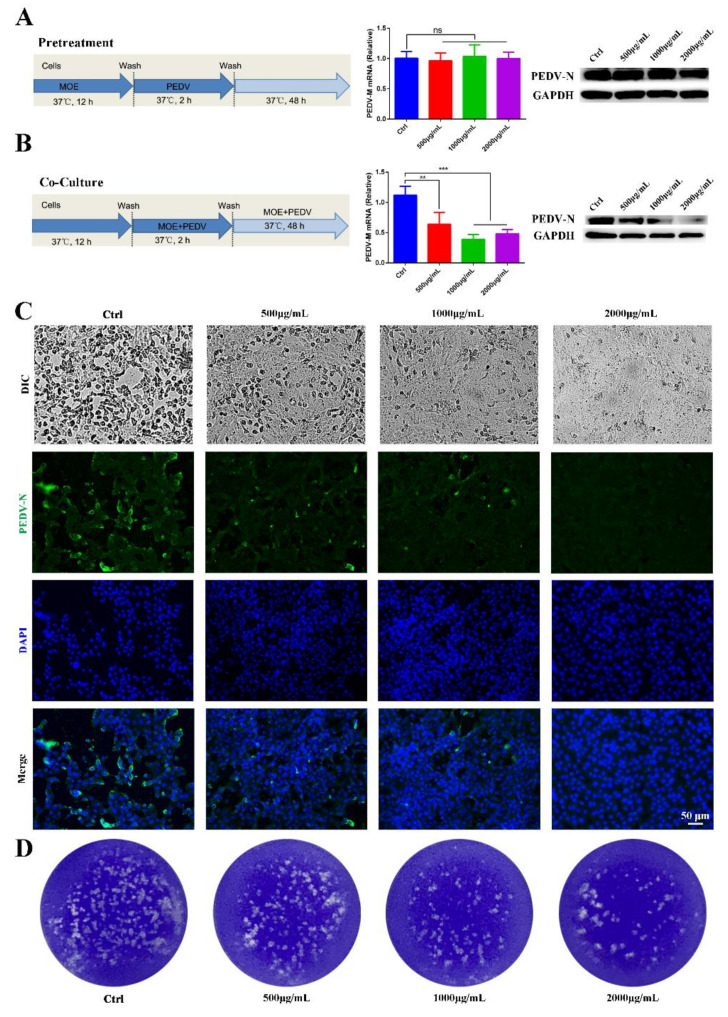
Figure 2.
Antiviral activity of MOE on PEDV infection on Vero cells. Vero cells were pretreated with MOE at different concentrations of 500, 1000, and 2000 μg/mL or PBS for 12 h, respectively, then the cells were infected with PEDV (MOI = 0.1) for 2 h. After extensive washing, cells were cultured with or without MOE for 48 h. (A) Schematic of time-of-addition analysis of MOE (500, 1000, and 2000 μg/mL) treatment against PEDV infection on Vero cells using pretreatment models, PEDV-M mRNA was detected by qRT-PCR, and PEDV-N protein was detected by Western blotting. (B) Co-culture models. PEDV-M mRNA was detected by using qRT-PCR, and PEDV-N protein was detected by Western blotting. (C) The light microscope of Vero cell to observe cytopathic effect. Viral yields were titrated by IFA. Green: PEDV-N; blue: DAPI; scale bars, 50 μm. (D) The virions were detected by plaque assay. ** p < 0.01; *** p < 0.001; ns, no significance. MOI: multiplicity of infection; MOE: the aqueous leaf extract of M. oleifera; PEDV: porcine epidemic diarrhea virus.

