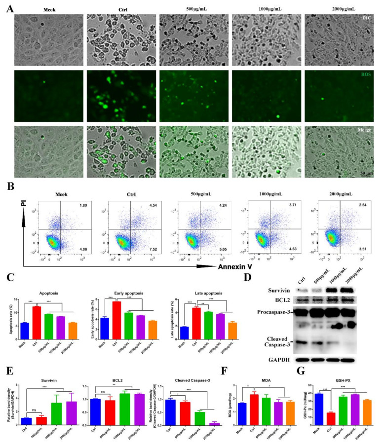
Figure 4.
MOE inhibited PEDV replication by suppressing PEDV induced apoptosis. (A) Vero cells were infected with PEDV (MOI = 0.1); after extensively washing, the medium was replaced with different concentrations of MOE for 48 h. Then, the cellular ROS was detected by DCFH-DA (10 μM, 20 min). (B) Apoptosis was quantified by combined staining with Annexin V and PI, and the fluorescence was analyzed at 36 h by flow cytometry. (C) Total apoptosis rate, early apoptosis, and late apoptosis rate were detected. Early apoptosis cells (Annexin V-positive); late apoptosis cell (positive for Annexin V and PI). Early apoptosis plus late apoptosis equals total apoptosis. (D) Survivin, BCL2, and Caspase-3 were detected by Western blotting. (E) The intensity of the bands (Survivin, BCL2, and cleaved Caspase-3) in terms of density was measured and normalized against GAPDH expression. (F) MDA production. (G) GSH-Px activity. Data are expressed as three independent experiments. * p < 0.05; ** p < 0.01; *** p < 0.001; ns, no significance. MOE: the aqueous leaf extract of M. oleifera; PEDV: porcine epidemic diarrhea virus; ROS: reactive oxygen species.

