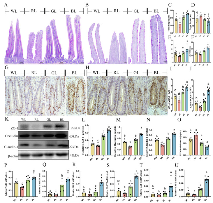
Figure 7.
Effects of different monochromatic lights on HE staining of jejunal tissue sections (A), PAS staining of jejunal tissue sections (B), jejunal villus height (scale bar = 100 μm) (C), jejunal crypt depth (D), jejunal villus height/crypt depth (V/C) ratio (E), goblet cell numbers (scale: 100 μm) (F), immunohistochemical staining photographs of MUC-2 (scale: 20 μm) (G), immunohistochemical staining photographs of PCNA (scale: 20 μm) (H), IOD of MUC2-positive cells (I), IOD of PCNA-positive cells (J), ZO-1, Claudin-1 and Occludin protein expression (K–N), intestinal permeability (O), PepT1 mRNA level (P), SI mRNA level (Q), SGLT1 mRNA level (R), GLUT2 mRNA level (S), CAT-1 mRNA level (T), CAT-2 mRNA level (U) in chicks of WL, RL, GL and BL at P42. WL: white light; RL: red light; GL: green light; BL: blue light. These results are shown as means ± SEM. Differences between the four groups are presented in the form of different letters (p < 0.05). PepT1, influx oligopeptide transporter peptide transporter 1; SI, sucrose-isomaltase; SGLT1, Na+-glucose cotransporter; GLUT2, glucose transporter type 2; CAT1, transporter 1 of cationic amino acid; CAT2, transporter 2 of cationic amino acid.

