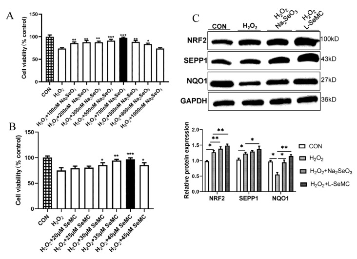
Figure 6.
Effects of Na2SeO3 and L-SeMC on oxidative stress in H2O2-induced AML-12 cells. (A) Cell viability of AML-12 cells was measured by CCK-8 after co-treatment with 100 μM H2O2 and different concentrations of Na2SeO3 (0–1000 nM) for 3 h (n = 7, *** p < 0.001, ** p < 0.01, * p < 0.05 vs. H2O2 group). (B) Cell viability of AML-12 cells was measured by CCK-8 after co-treatment with 100 μM H2O2 and different concentrations of L-SeMC (0–45 μM) for 3 h (n = 7, *** p < 0.001, ** p < 0.01, * p < 0.05 vs. H2O2 group). (C) The protein level of SEPP1, NRF2, and NQO1 (n = 7, ** p < 0.01, * p < 0.05).

