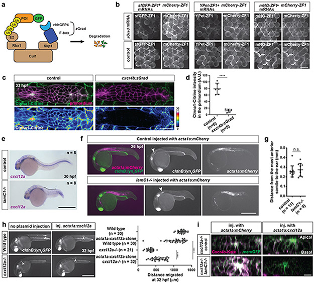
Extended Data Fig. 2. Depletion of Ctnna1-Citrine by zGrad and characterization of the lamC1 mutants.
a, Principle of zGrad-mediated protein degradation. b, Left: 8 hpf embryos injected with sfGFP-ZF1 mRNA and mCherry-ZF1 mRNA with or without co-injected zGrad mRNA. Middle: 8 hpf embryos injected with YPet-ZF1 mRNA and mCherry-ZF1 mRNA with or without co-injected zGrad mRNA. Right: 8 hpf embryos injected with mNeonGreen-ZF1 mRNA and mCherry-ZF1 mRNA with or without co-injected zGrad mRNA. n ≥ 20 embryos. Scale bar: 1 mm. c, Single confocal slices of primordia in prim:mem-mCherry; ctnna1:ctnna1-citrine control (left) and prim:mem-mCherry; ctnna1:ctnna1-citrine; cxcr4b:zGrad 32 hpf embryos (right). Lower panels show the Ctnna1-Citrine fluorescence as a heat map. Scale bar = 20 μm. d, Quantification of the Ctnna1-Citrine fluorescence intensity in control and zGrad-expressing embryos at 32 hpf. Data points, means, and SD are indicated. ****: p<0.0001 (two-tailed Welch’s t-test). e, Expression of cxcl12a in control (wild-type or lamc1−/+) and lamC1 mutant 30 hpf embryos. Bracket indicates the location of interrupted cxcl12a expression domain. Scale bar = 0.5 mm. f, mCherry-expressing clones in muscle of 26 hpf control (wild-type or lamC1−/+) and lamC1 mutant embryos also transgenic for cldnB:lyn2GFP. Arrowheads indicate the position of primordium. Scale bar = 0.5 mm. g, Quantification of the distance from the ear to the first somite in the indicated genotypes at 26 hpf. Data points, means, and SD are indicated. n.s.: p=0.5516 (two-tailed Mann-Whitney test). h, Images of the primordium in wild-type and cxcl12a−/− 32 hpf embryos with clones in the trunk muscle that express Cxcl12a together with mCherry (not shown) (Left). Asterisks indicate the ear and arrowheads the primordium. Scale bar = 0.5 mm. Quantification of the distance migrated by the primordium in the indicated experimental conditions at 32 hpf (Right). Data points, means, and SD are indicated. ****: p<0.0001 (One-way ANOVA followed by Tukey’s multiple comparison test). i, Cross-sectional images of the Cxcl12a sensor in primordia of cxcl12a−/− and cxcl12a−/−; lamC1−/− embryos with clones in the muscle of the trunk that express mCherry or Cxcl12a. Quantification shown in Fig. 2k. Scale bar = 20 μm. For d, e, g, h, n = number of embryos.

