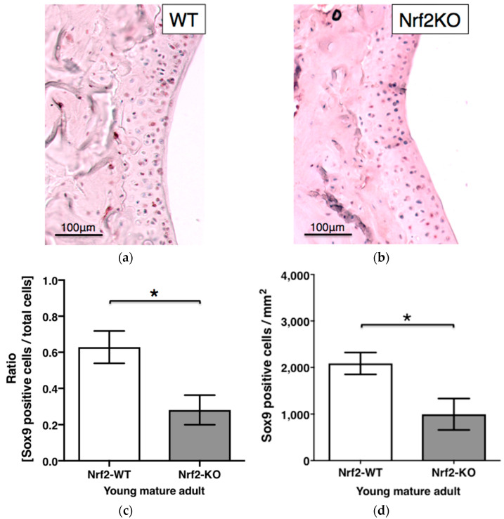
Figure 5.
Sox9 staining in hyaline articular cartilage of WT and Nrf2-KO mice. Representative immunohistochemistry of articular cartilage of a murine knee showing Sox9 staining in young mature adult WT (n = 5) (a) and Nrf2-KO (b) mice (n = 6). Sox9-positive articular chondrocytes were counted and expressed as ratios (c) and cell number per 1 mm2 area (d) in WT and Nrf2-KO mice, respectively. Plotted are means ± SEM. *, p < 0.05; WT, wild type; KO, knockout.

