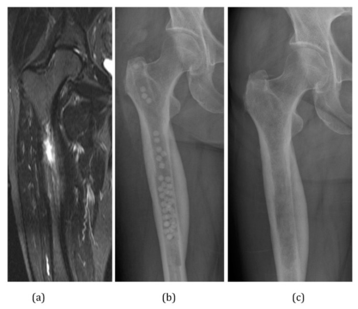
Figure 3.
(a) MRI image demonstrating extensive medullary edema, intramedullary abscess, and cortical involucrum. (b) Infection treated by excision via medullary reaming. Dead space filled with calcium sulphate pellets loaded with gentamicin. Shown via radiograph (c) follow-up radiograph at 4 months post-operatively. Calcium sulphate pellets have dissolved completely. Reprinted from Ferguson et al. [68] following the Creative Commons Attribution (CC BY) license (http://creativecommons.org/licenses/by/4.0/) (accessed on 16 September 2021).

