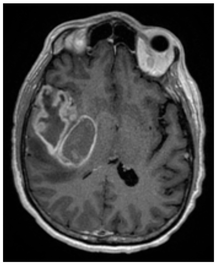
Figure 1.
T1 post-contrast axial MRI of a 60-year-old man who presented with a 3-month history of progressively worsening headaches and memory loss. The MRI was consistent with a large contrast-enhancing mass in the right temporal lobe. He underwent a gross total resection, which demonstrated a 1p19q intact, IDH-wildtype, WHO grade 4 glioma, MGMT promoter unmethylated. Surgery was followed by concurrent external beam radiation therapy (60 Gy in 30 fractions) with concurrent TMZ, followed by adjuvant TMZ and TTF. He succumbed to his disease at 13 months following diagnosis.

