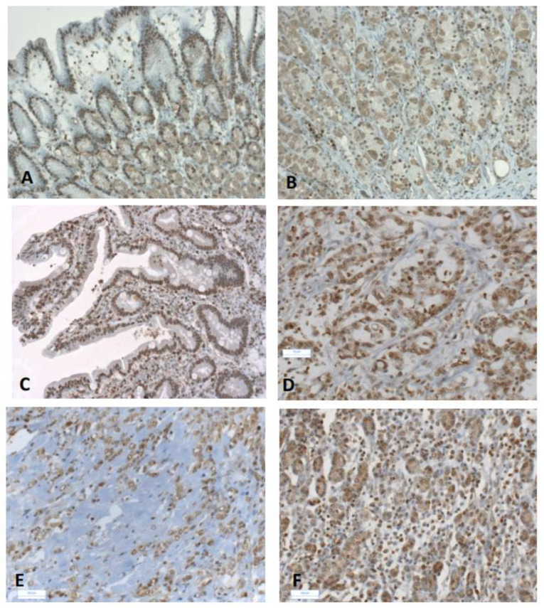
Figure 1.
Immunohistochemical staining of AhR in gastric cancers. Representative immunostaining of AhR in non-tumoral gastric mucosa (A,B) and in gastric cancers (C–F). Weak cytoplasmic and nuclear expression of AhR were observed in epithelial cells (A,B). Intestinal subtype GC with metaplasia (TNM 2a) (C); strong nuclear AhR staining in epithelial and stromal cells (C). Moderately differentiated intestinal subtype (TNM2a) showing nuclear AhR staining in tubular glands and stroma (D). Advanced diffuse GC (TNM4) (E): the intensity of AhR immunostaining was lower in the scattered cells of single ring cell component (SRCC). Early diffuse GC (TNM2a) (F): AhR immunostaining in epithelial and stromal cells. Original magnification ×10 (A,C,E); ×20 (B,D,F).

