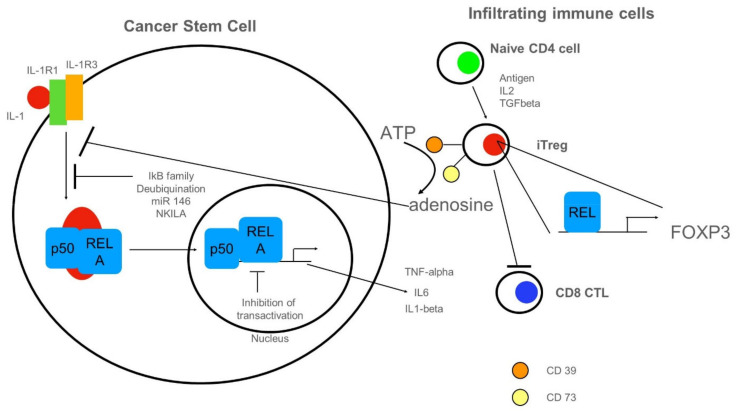
Figure 4.
Anti-inflammatory cues impinging on NF-κB in CSCs. On the left, a CSC with NF-κB-signaling is depicted. NF-κB could be activated by cancer-produced IL-1. Inhibition of NF-κB-activation could be mediated by several members of the IκB family, by the de-ubiquitination of signaling molecules (for details see text). Furthermore, non-coding RNAs such as microRNA(miR) 146 or long non-coding RNAs NKILA could inhibit NF-κB-activation by targeting activating proteins. Inhibition of transactivation could be driven by IEX-1 or by COMMD1-mediated ubiquitination of RELA. On the right side, tumor-infiltrating immune cells are shown. Naïve CD4+ cells can differentiate into induced regulatory T cells (iTregs). These express ectoenzymes such as CD39 and CD73 responsible for the hydrolysis of ATP to adenosine, in turn, inhibiting NF-κB-activation in CSCs. iTREGs could fancy cREL for expression of FOXP3 lineage transcription factor. A pro-tumor function of iTREGs could be the repression of CD8 cytotoxic lymphocytes (CTL).

