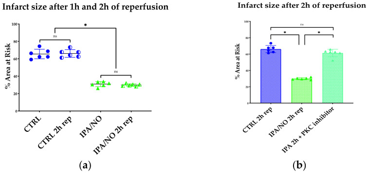
Figure 3.
(a) Infarct size expressed as a percentage of the area at risk (left ventricle mass). Neither the CTRL nor IPA/NO-treated heart showed a significant difference in terms of necrosis after 2 h of reperfusion when compared to 1 h. n = 6 for each experimental group. Data are expressed as Average ± Standard Deviation. * p < 0.001, ns: non-significant. (b) Infarct size expressed as a percentage of the area at risk (left ventricle mass). IPA/NO protection is abolished by co-infusion of a specific inhibitor of PKC translocation. N = 6 for each experimental group. Data are expressed as Average ± Standard Deviation. * p < 0.001, ns = p > 0.05.

