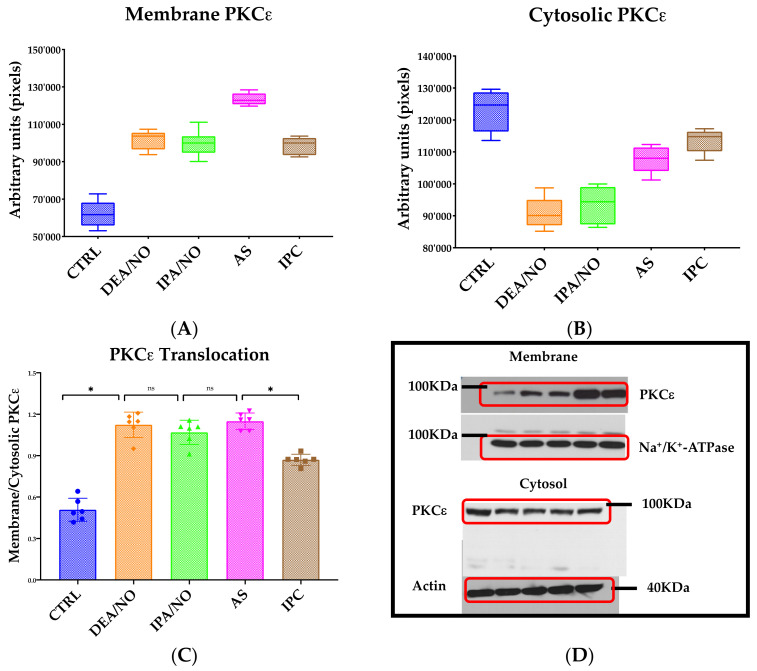
Figure 4.
(A) Quantification of membrane PKCε in myocardial tissue of ischemic hearts. Isolated hearts perfused with DEA/NO, IPA/NO, AS, or preconditioned with IPC. Membrane PKCε is higher in all treatments when compared to the CTRL group. All readings have been performed in triplicate maintaining the same ROI. (B) Quantification of cytosolic PKCε in myocardial tissue of ischemic hearts. Isolated hearts perfused with DEA/NO, IPA/NO, AS, or preconditioned with IPC. Cytosolic PKCε is lower in all treatments when compared to the CTRL group. All readings have been performed in triplicate maintaining the same ROI. (C) Ratio of membrane/cytosolic PKCε in myocardial tissue of ischemic hearts. Isolated hearts perfused with DEA/NO, IPA/NO, AS, or preconditioned with IPC. The ratio is significantly increased in all groups compared to the CTRL (p < 0.0001). All donors also show a significantly higher PKCε translocation when compared to IPC (* p < 0.001, ns = p > 0.05). (D) Representative images of Western blot membraned incubated with specific antibodies and revealed in chemiluminescence.

