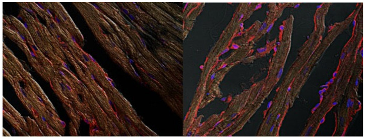
Figure 5.
Visualization of PKCε translocation induced by the novel compound IPA/NO. After treatment, hearts were processed to obtain thin slices to be probed with a PKCε antibody. Images were taken by confocal microscopy at 63×. In the left panel, a representative image on untreated, myocardial tissue, in the right panel a IPA/NO treated heart.

