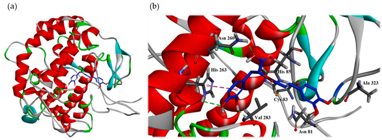
Figure 3.
Interaction of rosmarinic acid with the active sites of mushroom tyrosinase. The binding model (a) and the hydrophilic interactive model (b) of rosmarinic acid in the substrate binding pocket of the crystal structure (PDB: 2Y9X). The carbon atom of rosmarinic acid is in the dark blue color, the carbon atom of protein is in the blue color, the oxygen atom is in the red color and hydrogen atom is in the white color. The green line indicates the hydrogen bond interaction. The pink line indicates the π-π interaction.

