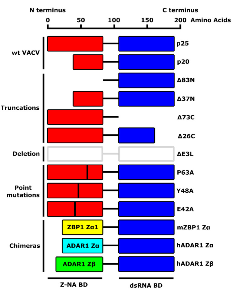
Figure 2.
A schematic diagram of VACV E3 protein and its mutants described in this review. VACV E3 protein comprises an N-terminal Z-nucleic acid (NA)-binding domain (BD) (also referred to as Zα) and a C-terminal dsRNA BD. Wild-type VACV E3 naturally occurs in two forms: p25 (full-length protein) and p20 (N-terminal truncation, resulting from leaky-scanning translation). Described mutants include truncations (E3LΔ83N, E3LΔ37N, E3LΔ73C, E3LΔ26C), a deletion of the entire E3 protein (ΔE3L), single amino acid substitutions (E3L P63A, E3L Y48A, E3L E42A), and chimeric E3 mutants with the Zα domain substituted with homologous Z-NA BDs from Z-DNA-binding protein 1 (ZBP1) or RNA-specific adenosine deaminase 1 (ADAR1).

