Figure 5.
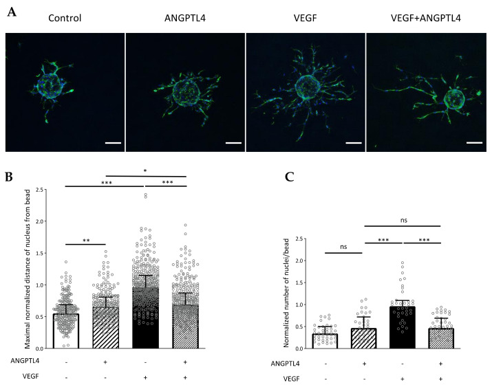
Regulation of 3D endothelial cell migration by ANGPTL4 and VEGF. (A) Cell responses were assessed by incubating endothelial cells seeded on beads with endothelial growth medium without or with ANGPTL4 (2.5 µg/mL) and/or VEGF (2.5 ng/mL) for 48 h. Cells were fixed and labelled for F-actin (green) and nuclei (blue). Scale bar: 100 µm. (B) Median values of normalized maximal distance covered by cell (nucleus) from the bead for each condition (+interquartile). (C) Median values of normalized number of nuclei emerging from a bead (+interquartile). Nuclei closely surrounding the bead are not considered. Individual values are normalized to the mean value of VEGF condition for each experiment. Values were measured in three independent experiments. ns: p > 0.05, * p ≤ 0.05, ** p ≤ 0.01, *** p ≤ 0.001 (Kruskal-Wallis).

