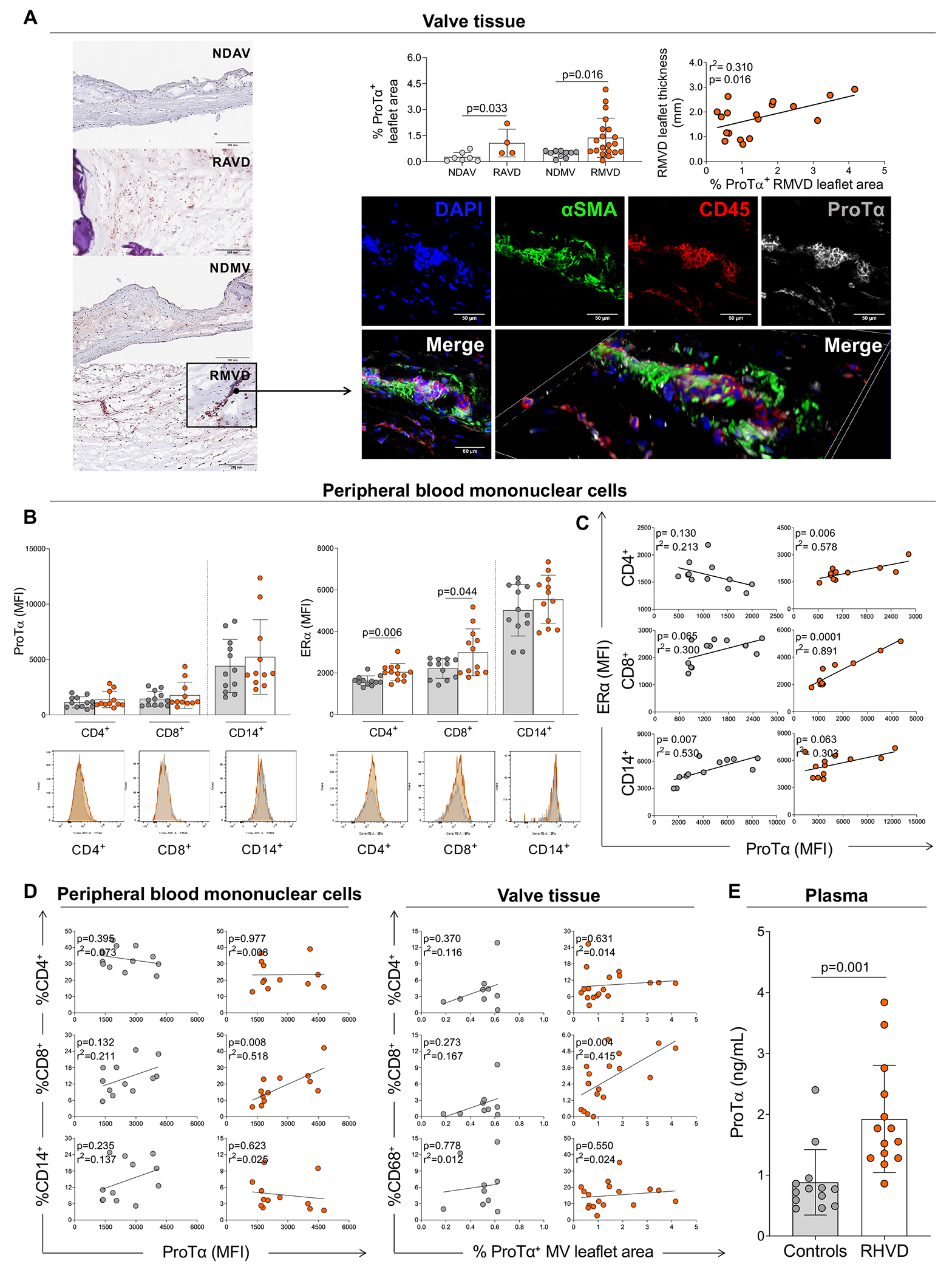
Figure 2: Ex vivo analysis of ProTα expression in heart valves and peripheral blood mononuclear cells in non-diseased and RHVD patients.

A, Histological evaluation of ProTα expression by immunohistochemistry staining in non-diseased aortic and mitral valves (NDAV, n=7 and NDMV, n=10) and rheumatic aortic and mitral valve disease (RAVD, n=4 and RMVD n=20). Quantification is reported as percentage of ProTα-positive area (% ProTα+ leaflet area) of the total leaflet area. Scale bars = 200 μm. Correlation analysis between RMVD leaflet thickness and ProTα+ RMVD leaflet area. Representative direct immunofluorescence staining for αSMA, CD45, and ProTα in RMVD. Scale bars = 50 μm. B, Median of intensity of fluorescence (MFI) of ProTα and estrogen receptor alpha (ERα) in healthy donors (gray dots) and rheumatic heart valve disease (RHVD) patients (orange dots) by different cell populations (CD4+, CD8+, and CD14+) in peripheral blood mononuclear cells. Representative histograms are show for each marker. C, Correlation analysis between MFI of ERα and ProTα by different cell populations (CD4+, CD8+, and CD14+) in peripheral blood mononuclear cells. D, Correlation analysis between percentage of CD4+, CD8+, and CD14+/CD68+ (valvular tissue/peripheral blood mononuclear cells) and ProTα. E, Plasma levels of ProTα in on health donors and RHVD patients. Bar graphs show the mean of values in each group and standard deviation. Two-group comparisons between non-diseased valves and disease valves were made using an unpaired t-test. Statistical significance is indicated in each graph.
