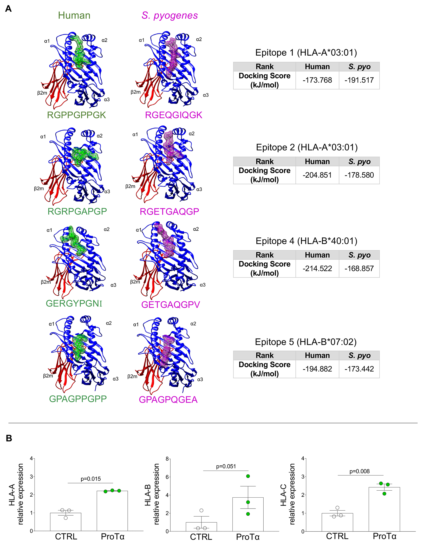
Figure 5: Peptide docking of type 1 collagen mimic epitopes and effects of recombinant ProTα on HLA-I expression by VICs.

A, Peptide docking of four identified human collagen type 1 mimic epitopes (green) and homologous region on S. pyogenes (magenta) sequences showing their interaction on HLA-I groove. Peptides are positioned between α1 and α2 HLA-I domains. Binding energy scores are shown for each comparison pair. B, mRNA expression levels of HLA-A, B and by VICs non-stimulated (CTRL, white dots, n=3) and stimulated with recombinant ProTα for 24 hours (green, n=3). Bar graphs show the mean gene expression relative to the CTRL culture with error bars representing standard deviation. Two-group comparisons between non-stimulated and stimulated cell cultures were made using a paired t-test. Statistical significance is indicated in each graph.
