Figure 1.
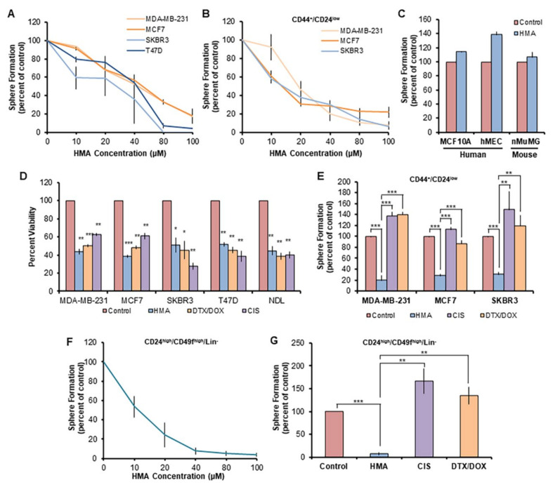
HMA ablates chemotherapy-resistant BCSCs but not normal mammary stem-like cells. (A) Secondary tumorsphere formation by human breast cancer cell lines was assessed after pretreatment with various concentrations of HMA for 24 h. Data are representative of three independent experiments. (B) Secondary sphere formation by CD44+/CD24low BCSC-enriched human breast cancer cells after HMA pretreatment. (C) Secondary mammosphere formation of non-transformed human and mouse mammary epithelial cell lines was assessed after 40 μM HMA treatment for 24 h. (D) Total populations of MDA-MB-231, MCF7, SKBR3, and T47D cells were treated with vehicle (DMSO), 40 μM HMA, 40 μM cisplatin (CIS), or a combination of 170 nM doxorubicin (DOX) and 50 nM docetaxel (DTX) for 24 h, and cell viability was determined by trypan blue exclusion assay. (E) Sorted human BCSC cells were treated with vehicle, 40 μM HMA, 40 μM CIS, or 170 nM DOX with 50 nM DTX, and secondary sphere formation was determined. (F,G) Cells dissociated from MMTV-NDL mouse mammary tumors were sorted (CD24high/CD49fhigh/Lin−) to enrich for the BCSC population, and 7-day sphere formation following pretreatment with HMA (F) or standard chemotherapeutics (G) was quantified over four biological tumor replicates from independent mice. Error bars represent SEM. *, p < 0.05; **, p < 0.01; ***, p < 0.001, by Student’s t-test.

