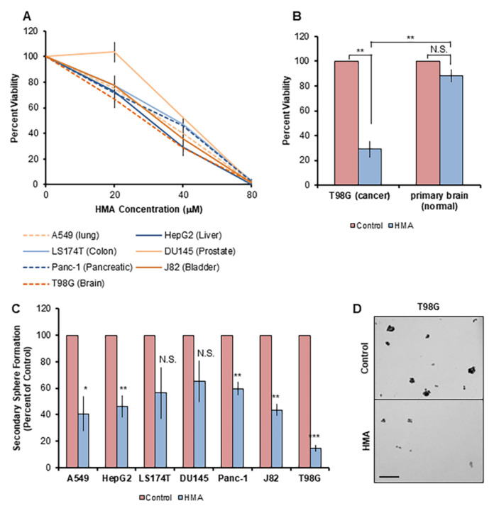
Figure 5.
HMA depletes CSCs derived from an array of human tissue types. (A) The viability of human tumor cell lines treated with varying concentrations of HMA for 24 h was assessed by trypan blue exclusion assay. Data are presented as averages of at least three independent biological trials and expressed as a percent of vehicle control. (B) T98G glioblastoma cells and non-transformed mouse primary brain glial cells were subjected to 24 h treatment with 40 μM HMA. Cell viability representative of three replicate trials is normalized to vehicle control. (C) Cell lines were treated with vehicle or 40 μM HMA for 24 h and then subjected to the sphere formation assay. Secondary sphere formation is presented as the average sphere count of at least three independent biological experiments and normalized to vehicle control. (D) Representative images of DMSO control and 40 μM HMA-treated T98G spheres are displayed. Scale bar = 200 μm. Error bars represent SEM. *, p < 0.05; **, p < 0.01; ***, p < 0.001, by Student’s t-test.

