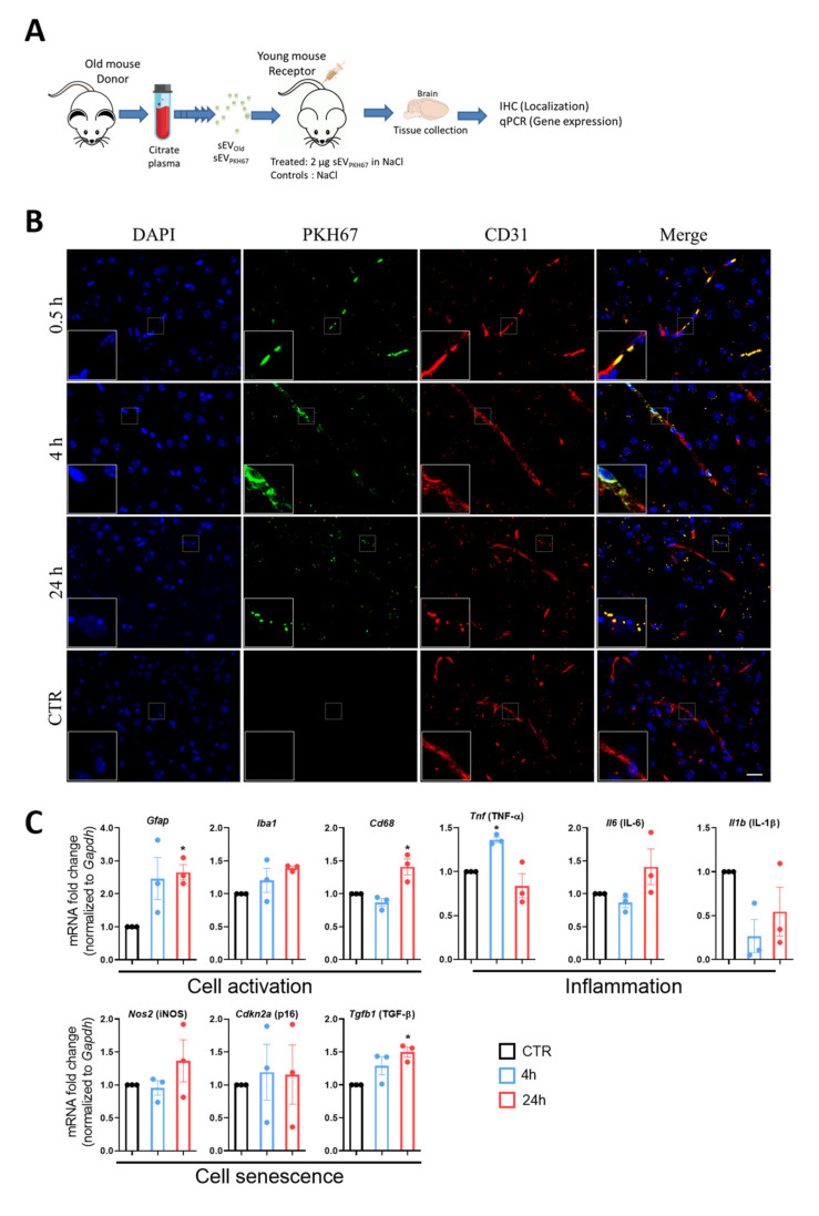
Figure 2.
Localization and effects of peripherally injected sEVs in vivo. (A) Scheme of sEV treatment. sEVs were isolated from plasma of 24-month-old donor mice and stained with PHK67. Two µg-labeled sEVs were injected into the tail veins of 3-month-old mice. Brains were collected at 0.5, 4, and 24 h. (B) The representative confocal microscopic images show exogenous-labeled sEVs localizing mostly in the vascular compartment of the brain (at 30 min) and in the parenchyma (at 4 and 24 h after injection). Platelet endothelial cell adhesion molecule-1 (CD31) was stained with rhodamine (red) and nuclei were stained with DAPI (blue) for visualization. Dashed boxes are shown at 3X magnification at the bottom left of each image. Scale bar 20 µm. (C) Time-dependent effect of exogenous sEVs on gene expression in young mice. Gene expression was assessed 4 (blue bars and dots) and 24 h (red bars and dots) after treatment by PCR normalized to Gapdh and compared to the respective controls (black bars and dots) (CTR; set to 1.0). For display purposes, only one bar is presented. Results are shown as mean ± SEM. * p < 0.05 against CTR. One-way ANOVA with Dunnett’s multiple comparisons test.

