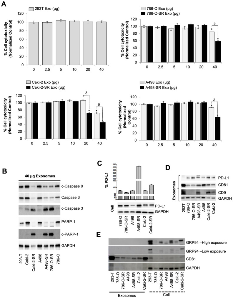
Figure 1.
SR RCC exosome’s role in suppressing the immune system. (A) Effect of isolated exosomes from 293T or various RCC cells on Jurkat T cell proliferation determined by WST-8 assay after 72 h incubation (n = 4). (B) Caspase activation in Jurkat T cells after dosing with 40 µg/mL exosomes for 72 h. Caspases 3, 9, and PAPR-1 were then assessed using western blotting. GAPDH was used as an internal control. Full blots are presented in Supplementary Figure S8. (C) PD-L1 protein in ccRCC cell lines was detected by western blotting. Full blots are presented in Figure S5. (D) Western blot analysis of the expression of exosomal PD-L1, CD9, CD81, and GAPDH in 293T, or SS, or SR RCC cell lines. (E) Western blot analysis of exosome and cell lysates identifying the presence of GRP94, a marker specific to large EVs, only in the latter. Full blots are presented in Supplementary Figure S8. * p < 0.05, and δ p < 0.01.

