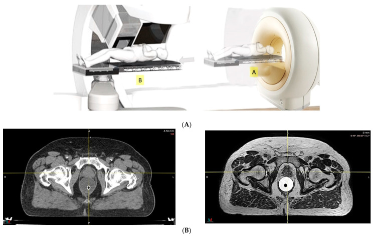
Figure 3.
(A) Diagram of a proton therapy system with in-room magnetic resonance (MR). MR images are acquired first, with the patient lying on a robotic couch in position A, followed by relocation to position B for proton delivery, directly after image registration. (B) Comparison of computed tomography and MR images of prostate cancer.

