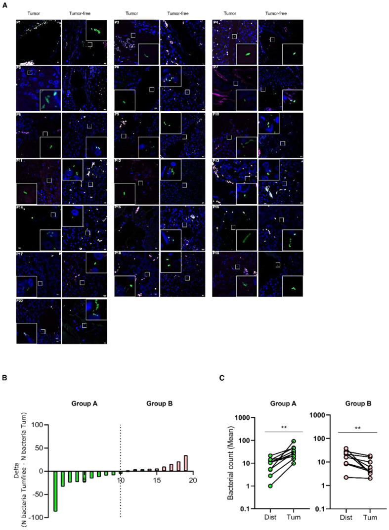
Figure 3.
Comparison between bacterial infiltration in paired tumoral and tumor-free pan-NEN tissue. (A) Representative confocal microscopy images of the bacterial signal observed within paired tumoral and tumor-free pan-NEN patients (N = 18). Scale bar = 10 µm. (B) Bar plot representing the difference between bacteria abundance within paired tumoral and tumor-free tissue (N bacteria Tumfree-N bacteriaTum) (N = 19). (C) Comparison between the mean of bacterial count within paired tumoral and tumor-free tissue of Group A and B. Wilcoxon test was used to assess statistical significance. ** p-value < 0.05.

