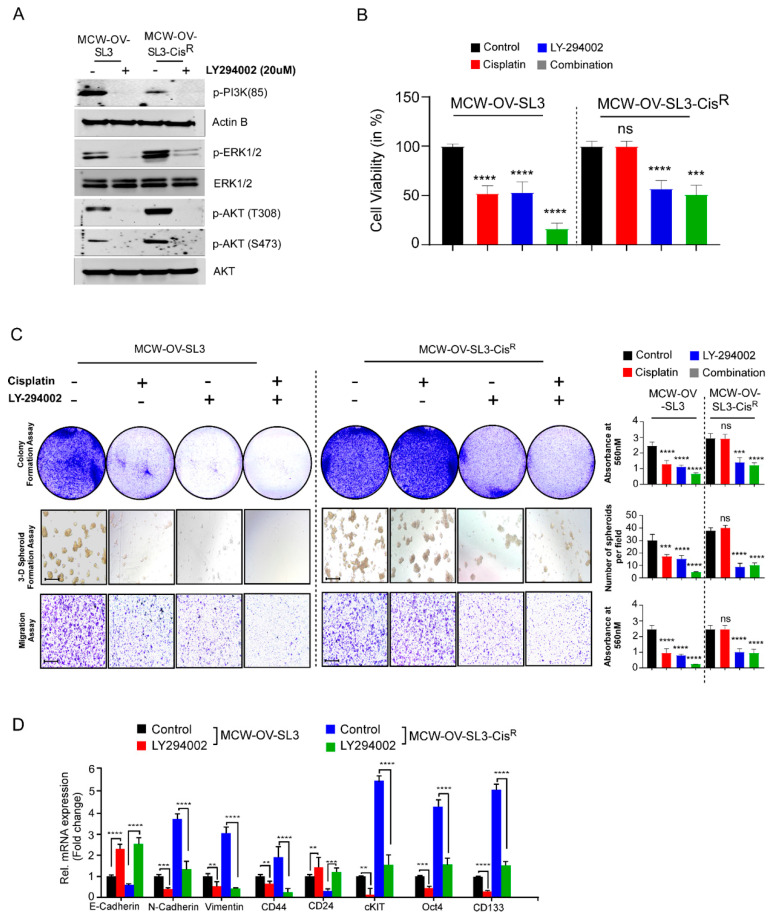
Figure 5.
PI3K inhibitor treatment reduced EMT, cancer stemness, and chemoresistance in the MCW-OV-SL3 cell line. (A) MCW-OV-SL3 and MCW-OV-SL3-CisR cells were treated for 24 h with LY-294002. Cell lysates were prepared and immunoblotted using the antibodies indicated (see Figure S3). (B) MCW-OV-SL3 and MCW-OV-SL3-CisR cells were treated with either cisplatin (2.2 and 6.2 µM), LY-294002 (5 and 10 µM), or their combination (cisplatin 1.1 and 3.1 µM), LY-294002 (2.5 and 5 µM) for 48 h and the cell viability was assessed by MTT assay at 48 h. Results are representative of three independent experiments performed in triplicates and error bars represent SE of the mean. (C) MCW-OV-SL-3 and MCW-OV-SL-3-CisR cell lines treated for 48 h with either cisplatin, LY294002, or in combination (as treated in B) were grown on 6-well culture plates for 14 days for colony formation, then stained using 0.5% crystal violet and photographed. For 3D formation assay, cells were grown on Matrigel-coated plates for seven days and then photographed using a phase-contrast microscope. For cell migration assay, cells were plated on trans-well inserts without Matrigel for 12 h. Migrated cells were photographed (left panel). Scale bars represent 500 µm. The right bar graph reveals the quantification of the colony formation, 3D spheroids, and migratory ability of indicated cell lines. (D) Total RNA was isolated, and qPCR was performed from MCW-OV-SL-3 and MCW-OV-SL-3-CisR cell lines treated with either cisplatin, LY294002, or in combination (as treated in B) for 48 h. mRNA expression was normalized to GAPDH. Bars represent SE of triplicate determinations. Error bar indicates SE of triplicate determination. ns-not significant, **p < 0.01, *** p < 0.001, **** p < 0.0001 statistically significant compared with control.

