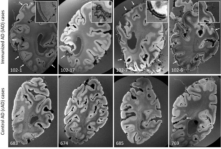
Figure 1.
Ex vivo GRE 7T MRI reveals haemorrhagic lesions in iAD, but not cAD cases. Top row shows examples of curvilinear cortical hypointensities (arrows) that were observed on ex vivo GRE 7T MRI scans of formalin-fixed coronal slices in five out of 10 iAD cases, representative of cortical superficial siderosis. Similar abnormalities were not observed in cAD cases (bottom row), except for a small area of cortical hypointensity in one case (arrow). Note the appearance of confluent hypointense abnormalities in the white matter in case 102-6, which may reflect tissue rarefaction related to formalin fixation, and was observed in 4 out of 10 iAD cases, but not in the cAD cases.

