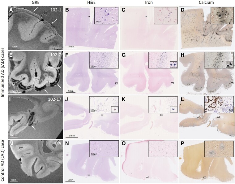
Figure 2.
Observed haemorrhagic lesions on ex vivo MRI correspond to iron and calcium deposits on histopathology. An area with curvilinear cortical hypointensities (arrows) on ex vivo GRE 7T MRI in positive control iAD Case #1 (A) revealed on histopathology tissue abnormalities in the cortex on H&E (B), which were confirmed as intracellular iron-positive depositions on an adjacent Perls’ Prussian blue-stained section (C) and intracellular calcium depositions on an adjacent Von Kossa-stained section (D). In another iAD case with a more severe degree of cortical hypointensities (E, arrows), the corresponding histopathological sections revealed more extensive tissue abnormalities (F), in the form of intracellular iron depositions (G) and intracellular calcium depositions (H). In another iAD case with focal cortical hypointensity (I, arrow), the corresponding histopathological sections revealed haemosiderin deposits at the level of the leptomeningeal vessels (J), which stained positive for iron (K) and calcium (L). The inset in (L) shows extensive leptomeningeal CAA as well as concentric vessel wall splitting as observed on an adjacent section that underwent immunohistochemistry against Aβ. The bottom row shows a representative example of one of the cAD cases, without abnormalities on ex vivo GRE 7T MRI (M), H&E (N), Perls (O) or Von Kossa-stained sections (P). Note the presence of extracellular calcium depositions in (D, H, L, P), which may be the result of prolonged formalin fixation of banked tissue.

