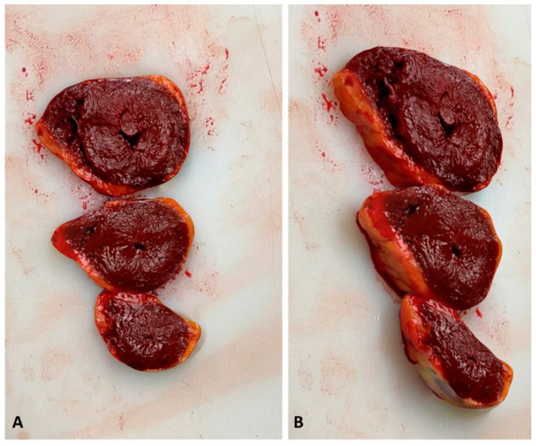
Figure 5.
Three-dimensional reconstruction of the heart slides performed at the macroscopic examination of the heart (case 3). (A) shows a perpendicular view of the 3D model. (B) shows a lateral view. The myocardial wall is precisely measurable in the 3D model, sustaining the hypothesis of myocardial hypertrophy. In this case, the histologic examination confirmed the diagnosis.

