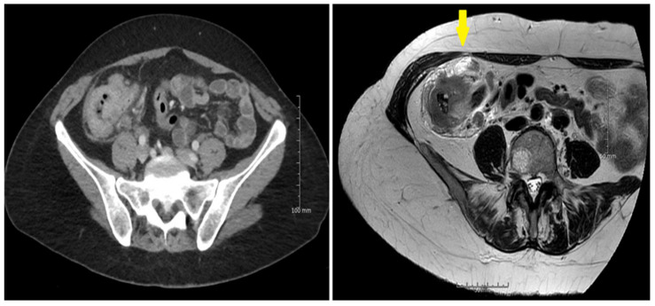
Figure 3.
(left) The serosal involvement of a large cecal pT4a,N1,V2 tumor was not identified by CT. (right) MRI identified the serosal involvement seen at the anterior part of the tumor (arrow). MRI also spotted lymph node metastases as well as extra mural vascular involvement, confirmed at the histopathological examination.

