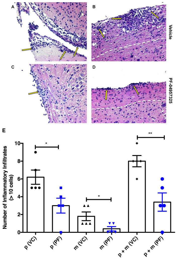
Figure 2.
Treatment with PF-04957325 suppresses histopathologic signs of EAE in the CNS. Mice with an EAE score of at least 1 were treated with a vehicle control in an equivalent volume of DMSO and PBS to the corresponding inhibitor dose or PF-04957325 from day 10 to day 13 (twice daily for 4 days), followed by the dissection of their brains and spinal cords for histopathological analysis. The sections were stained with HE, and their inflammatory foci were quantitated according to the published protocols [35,45]. Images of the brain (A,C) and spinal cord (B,D) sections of the vehicle control (grade 4) and PF-04957325 (grade 2)-treated mice are shown (original magnification 20X). The arrows point to inflammatory foci, and the white lines indicate the border between white and grey matter in the spinal cord. (E) The graph represents the number of inflammatory foci containing >10 cells [35,45] present in the spinal cord parenchyma (p) (* p = 0.025), brain meninges (m) (* p = 0.034), and spinal cord parenchyma plus brain meninges (p + m) (** p = 0.005), as assessed in HE sections of the vehicle control (V)- and PF-04957325 (PF)-treated mice. (n = five mice per group).

