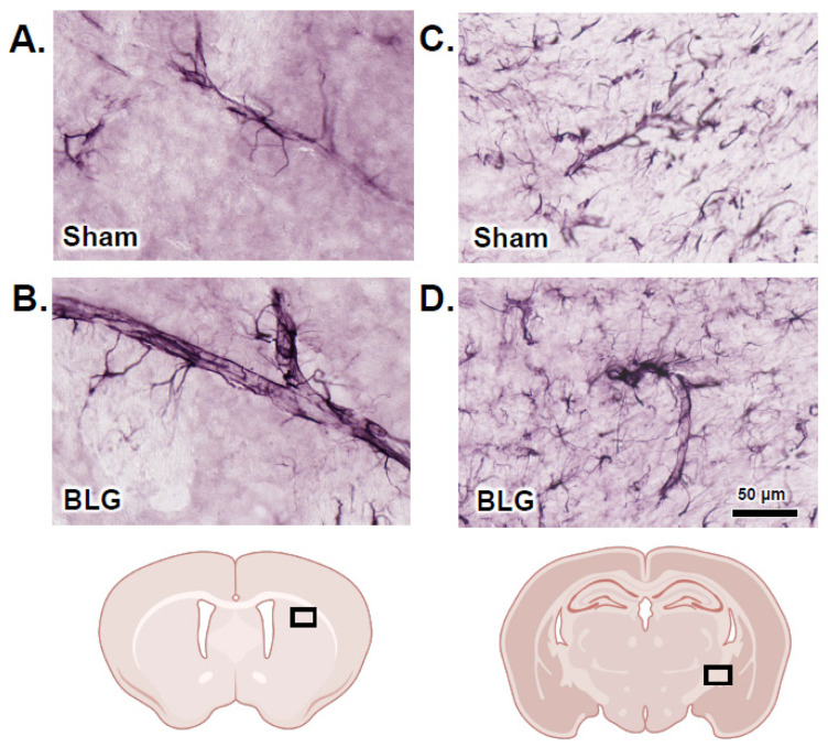
Figure 12.
Immunohistochemical staining of glial fibrillary acidic protein (GFAP)-positive perivascular astrocytes. Brain sections from the sham (A,C) and BLG-sensitized (B,D) mice were subjected to immunohistochemical staining for GFAP to identify astrocytes. Photomicrographs of GFAP-immunoreactive perivascular astrocytes were taken from the striatum (A,B) and the internal capsule (C,D) with a 40× objective. The rectangles in the brain diagrams indicate the approximate locations where the photomicrographs were taken. Scale bar: 50 µm (for all panels).

