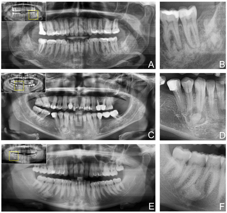
Figure 2.
Study participants’ two-dimensional panoramic radiography (PAN) providing anatomical information of the fibro-osseous lesion. (A) shows an ossifying fibroma in the posterior mandible. In contrast (C) shows cemento-osseous dysplasia at the canine in the fourth quadrant, while (E) shows a cemento-osseous dysplasia at the first molar in the fourth quadrant. For orientation, the dotted rectangles in the corner show the enlarged area of the region of interest (B,D,F).

