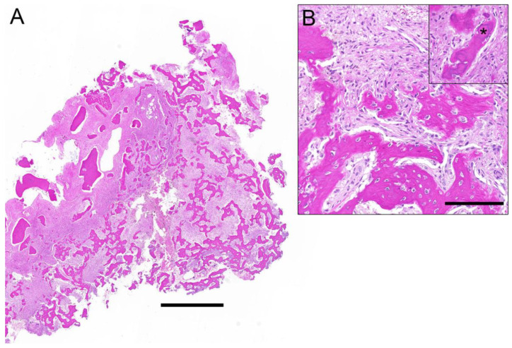
Figure 3.
Histological overview of a fibro-osseous lesion (A) depicts irregular bony trabeculae with different grades of maturation and a fibroblastic spindle cell proliferation in between. The magnification (B) shows the bony islands with lack of osteoblastic rimming and the interjacent bland spindle cell proliferation. Inset illustrates the small spheroid cementum-like particles (asterisk). Correlation with radiological images rendered a diagnosis of cemento-osseous dysplasia. Scale bar 1 mm (A) and 100 µm (B).

