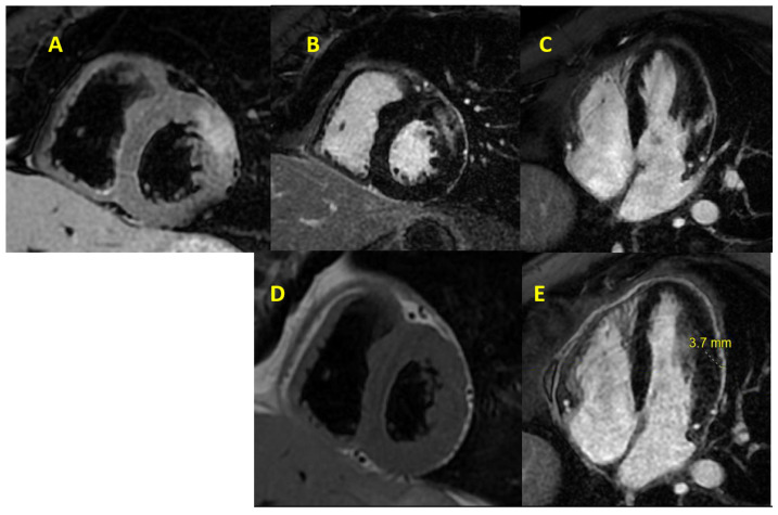
Figure 3.
A case of myocarditis that was diagnosed using CMR based on the 2018 revised LLC. (A): T2-weighted image (T2 double inversion non-contrast) showing edema in the mid-myocardial segment of the anterolateral wall (non-ischemic distribution). (B) Short-axis and (C) 4-chamber view LGE images showing high signal intensity (SI) in a non-ischemic distribution in the midwall along with pericardial involvement on LGE imaging. LGE imaging taken months after treatment as seen in (D,E) when compared to (B,C), respectively, showing no myocardial scar seen on LGE with only evidence of pericardial fibrosis. Image courtesy of Neeraja Yedlapati, MD, FACC, FASE, FSCCT.

