Figure 2.
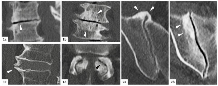
Imaging examples. (1): Degenerative lesions of the lumbar spine; (1a) = sagittal reconstruction: white arrowhead marks discal vacuum phenomenon and narrowing of intervertebral space; (1b) = coronal reconstruction: white arrowheads mark sclerosis of endplates; (1c) = sagittal reconstruction: white arrowhead indicates spondylophyte; (1d) = axial reconstruction: black arrowhead marks intraarticular vacuum phenomenon. Additionally, note the extensive sclerosis and joint space irregularities from OA of the facet joints. (2): degenerative lesions of the SIJ; (2a) = axial reconstruction: ventrally located, bridging osteophytes of the right SIJ marked with white arrowheads; (2b) = oblique-coronal reconstruction: extensive sclerosis around the joint (white arrowheads).

