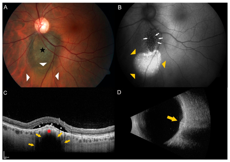
Figure 1.
Example of a juxtapapillary indeterminate choroidal melanocytic lesion showing two risk factors. (A) Color fundus photography of the left eye of a 35-year old female patient showing a pigmented choroidal lesion (black star) adjacent to the optic disc. Please note the presence of subretinal fluid over and inferior to the lesion (white triangles). (B) Autofluorescence imaging highlights the subretinal fluid (yellow triangles) and shows the presence of orange pigment (white arrows) over the lesion. (C) Enhanced depth spectral-domain optical coherence tomography (EDI-OCT) of the dome-shaped tumor (red star) shows choriocapillaris compression and choroidal shadowing (yellow arrows) as well as subretinal fluid (white stars) over the tumor. (D) B-scan ultrasonography shows a dense, dome-shaped lesion (yellow arrow) measuring 1.5 mm in thickness.

