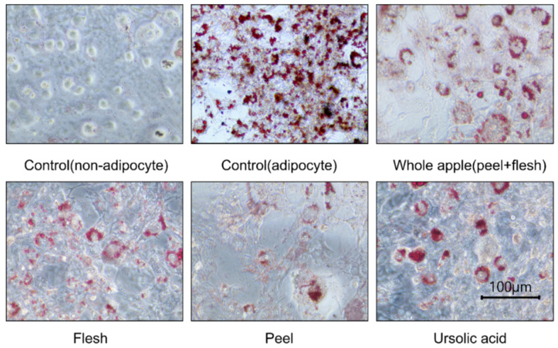
Figure 3.
3T3-L1 cell adipocyte images, observed and photographed using a microscope (×400) after cellular lipid accumulation was stained with Oil-Red O solution. The cells were from 70% methanol-treated extracts (1 μg dry powder in 1 mL of DMSO) of peel, flesh, and whole apple (peel + flesh) concentrated samples, or those treated with ursolic acid 5 μM. During the three repetitions of the treatments, a representative photograph was taken.

