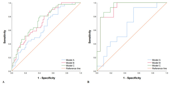
Figure 4.
Receiver operating characteristic (ROC) curves in both the training and the validation set. Method A, Method B, and Method C represented the modeling methods of clinical features (HDL + Apo B), ultrasound features (plaque volume + Rad-score), and the combined model (HDL + Apo B + plaque volume + Rad-score). (A) Training set: the AUCs in Model A, Model B, and Model C were 0.648 (95% CI, 0.543–0.753), 0.723 (95% CI, 0.627–0.818) and 0.741 (95% CI, 0.646–0.835), respectively. (B) Validation set: the AUCs in Model A, Model B, and Model C were 0.667 (95% CI, 0.485–0.848), 0.922 (95% CI, 0.833–1.000) and 0.939 (95% CI, 0.860–1.000), respectively.

