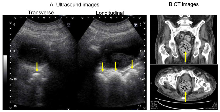
Figure 5.
Ultrasound images of a patient who had frequent episodes of diarrhoea with intense faecal impaction in the rectum on CT imaging. (A) Rectal ultrasound images showing a hyperechoic area indicating faecal retention (arrows). (B) CT image showing faecal retention in the rectum (arrows). The upper image shows a longitudinal section, and the lower image shows a transverse section. CT, computed tomography.

