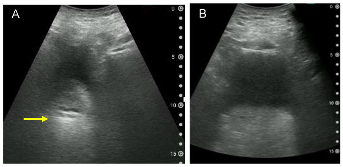
Figure 6.
Examples of a blurred ultrasound image of the bladder and rectum. (A) Blurred transverse ultrasound image of the rectum due to little urine in the bladder and intestinal gas. A hyperechoic area is observed, possibly indicating faecal retention (arrow). (B) Transverse ultrasound image of the rectum in an adult male with a body mass index of 28.6 kg/m2. Due to the thickness of the abdominal wall, the bladder and rectum cannot be clearly observed. The volume of urine voided immediately after imaging is approximately 180 mL.

