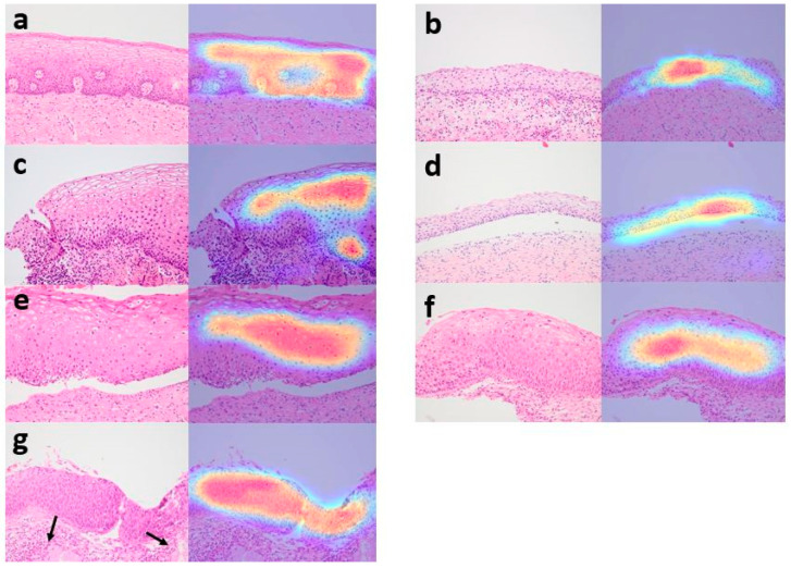
Figure 5.
Grad-CAM images by EfficientNet-B7. Normal squamous epithelium was highlighted in Grad-CAM images (a–d). Images from cervix interpreted as non-neoplasm by the EfficientNet-B7 include exocervix (a), metaplastic muco-sa from transformation zone (b), cervicitis and erosion (c) and atrophic mucosa (d). In CIN1, layers with koilocytotic cells were mainly highlighted (e). The highlighted areas extended to the upper two-third of the epithelium in CIN2 (f) and full-thickness of the epithelium in CIN3 (g). Normal endocervical glands ((g), black arrows) were not highlighted.

