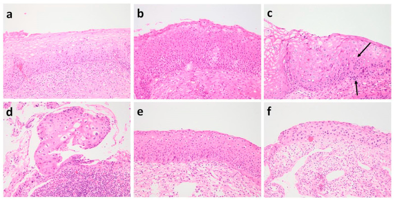
Figure A1.
Histology of misclassified cases by CNN models. A case with scarce koilocytotic cells but basal atypia was false-negative (a). CIN3 showing basal/parabasal-type atypia throughout most of the epithelium but not all was downgraded to CIN2 (b). CIN2 (c) downgraded as CIN1 showed koilocytotic changes in the upper half and maturation in upper most layers but had atypia focally extending to the lower half of the epithelium (black arrow). In CIN1 upgraded as CIN2, the epithelium was disoriented (d). CIN2 with koilocytosis (e) and atrophic CIN2 (f) were upgraded as CIN3.

