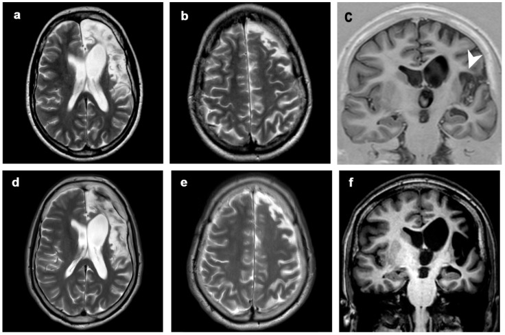
Figure 4.
Patient 2, progression of the disease in 21 years. Axial (a,b,d,e) T2-weighted images, coronal T1-inversion recovery (c) and T1-weighted image (f) at (a–c) 5 years (first MRI scan) and (d–f) 21 years from clinical onset (last MRI scan). The disease showed mild progression over time, involving left perisylvian region (including frontal, insular and temporal structures, arrowhead). Clinical symptoms progressed from seizures at onset to seizures, focal deficits, EPC, cognitive and behavioral disorders.

