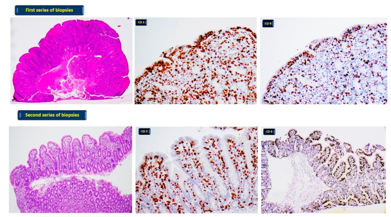
Figure 1.
First series of biopsies: patient without GFD; diffuse moderate-severe atrophy of villi (haematoxylin-eosin 40×) with pathological increase of T lymphocytes CD3 (400×) and CD8 positive (400×). Second series of biopsies: patient on GFD; normal villi with focal low atrophy (haematoxylin- eosin 100×) and pathological increase of T lymphocytes CD3 (400×) and CD8 positive (100×).

