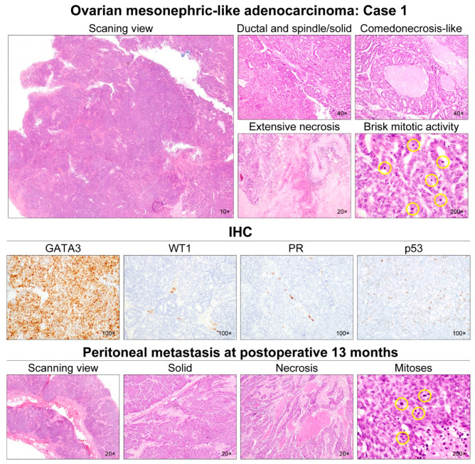
Figure 2.
Histological features of ovarian MLA: Case 1. The ovarian mass was a 7.5-cm cystic lesion with some internal mural nodules. At scanning magnification, the mural nodules appeared solid and deeply basophilic due to hypercellularity. They consisted of adenocarcinoma, showing ductal and solid growth patterns. Most of the tumor cells formed a complex cribriform and solid architecture with slit-like glandular spaces. We noted some foci of intraluminal necrosis and karyorrhexis within the cribriform architecture, resembling comedonecrosis observed in ductal carcinoma in situ of the breast. Even though most of the tumor cells exhibited mild-to-moderate nuclear pleomorphism, some areas showed severe nuclear pleomorphism, extensive coagulative tumor cell necrosis, and brisk mitotic activity (27/10 high-power fields). Immunostaining revealed that the tumor cells were completely negative for WT1, ER, PR, and p16; p53 expression pattern was wild-type. Since the tumor was confined within the right ovary (FIGO stage IA), the patient received no further treatment. However, at 13 months post-operatively, she developed multiple metastases in the abdominopelvic peritoneum, liver, and lungs. The metastatic lesions displayed solid growth pattern with scattered foci of coagulative tumor necrosis. Similar to the primary tumor, the metastatic tumor cells showed severe nuclear pleomorphism. Mitotic count (25/10 high-power fields) was similar to that of the primary tumor. The yellow circles are intended to highlight the mitotic figures, which just appear very small dark spots.

