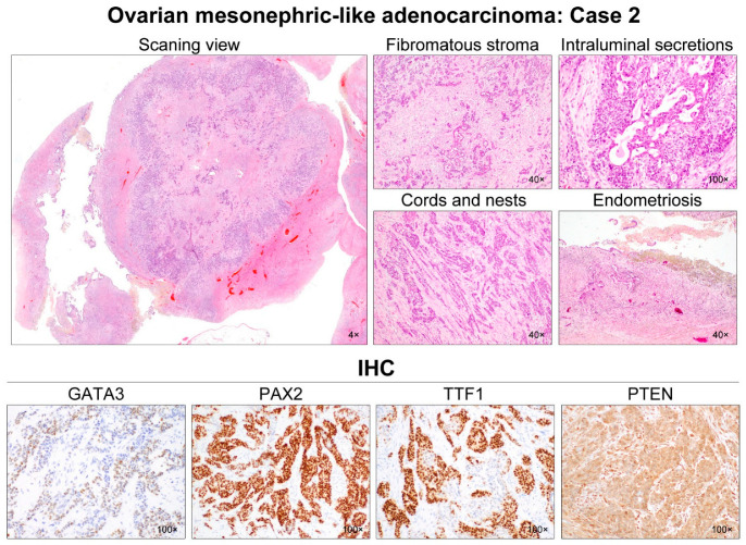
Figure 3.
Histological features of ovarian MLA: Case 2. A 4.7-cm ovarian mass contained small, nodular components confined to the cyst. The mural nodules consisted of solid tumor tissue showing infiltrating small tubules in the background of the fibromatous stroma. These tubules were haphazardly arranged and anastomosed with each other, forming a cribriform or trabecular architecture. In addition to the small tubular pattern, the tumor cells formed irregular-shaped nests and cords, separated by thin intervening fibrous stroma. Hyaline-like eosinophilic secretions were occasionally noted within the tubular lumina and dilated duct-like structures. No severe nuclear pleomorphism was identified. The cystic lining was primarily composed of endometrial-like glands and stroma, the latter of which was occupied by hemosiderin-laden macrophages. Immunohistochemically, the tumor cells were focally positive for GATA3 with moderate staining intensity. PAX2 immunoreactivity was uniform and intense throughout the tumor. TTF1 expression was also diffuse and strong in most of the tumor cells. Hormone receptor expressions were absent, and loss of PTEN expression was not observed.

