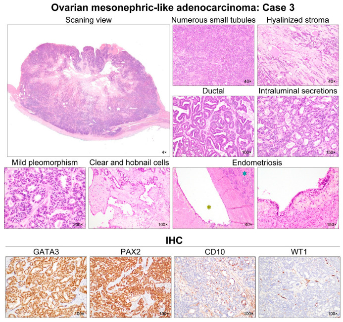
Figure 4.
Histological features of ovarian MLA: Case 3. Scanning magnification revealed that the solid component was a hypercellular tumor, with areas of cystic change and degeneration in the central portion. Numerous small tubules were compactly aggregated and merged. The amount of intervening stroma was small, but the central portion had some microscopic areas of stromal degeneration and hyaline fibrosis. An endometrioid-like glandular structure (i.e., ductal growth pattern), accompanied with focal maze-like architecture, comprised approximately one-third of the entire tumor volume. These tubules and endometrioid-like glands were lined by cuboidal and columnar epithelial cells, respectively, and possessed pale or dense eosinophilic intraluminal secretions. Most tumor cell nuclei demonstrated intermediate-grade atypia characterized by mild-to-moderate pleomorphism and hyperchromasia, with rare mitotic figures. In a few foci, dilated glands were lined by a single layer of tumor cells with clear-to-eosinophilic cytoplasm and a hobnail appearance. Their nuclei were similar in size and shape to those of the adjacent tubules. The intervening stroma showed sparse inflammatory infiltrate, and focal myxoid and hyaline degeneration. An endometriotic cyst (yellow asterisk) was observed adjacent to the tumor tissue (blue asterisk). The lining epithelium of the endometriotic cyst underwent extensive tubal metaplasia. The endometrial stroma was filled with hemosiderin-laden macrophages. Immunostaining revealed that the tumor cells were diffusely and strongly positive for GATA3 and PAX2. CD10 expression, along the luminal surface of the tubules and ducts, was characteristic of MLA. Lack of WT1 expression excluded the possibility of serous carcinoma.

