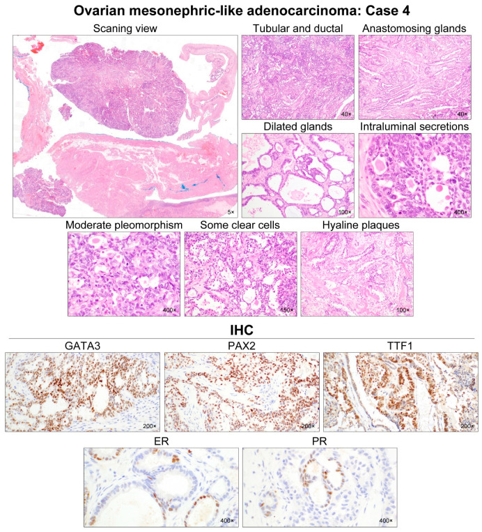
Figure 5.
Histological features of ovarian MLA: Case 4. The ovarian mass was solid and cystic, with mural nodules protruding into the cystic lumen. The cystic wall consisted of fibromuscular connective tissue, whereas the solid mural nodules were composed of adenocarcinoma. Two predominant growth patterns were tubular and ductal. Small tubules were compactly aggregated and admixed with inter-anastomosing glands. Cystically dilated glands were often seen. Hyaline-like intraluminal substances were frequently identified in the tubules and glands. The tumor cell nuclei exhibited mild-to-moderate pleomorphism. In addition, angulated ductal structures traversed by thick bands of hyaline collagen were observed. In a few foci, some tubules lined by a few layers of cuboidal or hobnail cells with clear cytoplasm raised the possibility of clear cell carcinoma (CCC). However, severe nuclear pleomorphism, prominent nucleoli, and myxoid stromal cores, characteristic of CCC, were absent. The tumor cells were strongly positive for GATA3, PAX2, and TTF1. We noted that some tumor cells were positive for ER and PR, with moderate staining intensity. Although hormone receptor immunoreactivities were not typical of MLA, previous data indicated that this unusual expression pattern did not preclude the diagnosis of MLA.

