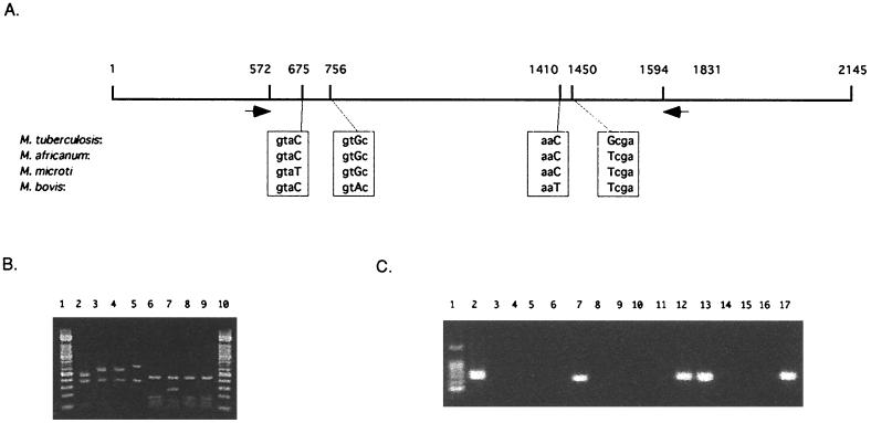
FIG. 3.
Differentiation of members of the M. tuberculosis complex by using PCR and PCR-RFLP. (A) gyrB sequences of the members of the M. tuberculosis complex. The sequences of polymorphic loci are enclosed in boxes, and capital letters indicate substituted sequences. Black arrows show the positions of primers for the PCR-RFLP analysis. (B) PCR-RFLP patterns obtained with the type strains of the M. tuberculosis complex. Lanes: 1 and 10, 100-bp ladder molecular size markers; 2 to 9, RFLP patterns of PCR products from M. microti (lanes 2 and 6), M. tuberculosis (lanes 3 and 7), M. africanum (lanes 4 and 8), and M. bovis (lanes 5 and 9). Lanes 2, 3, 4, and 5 show RFLP patterns obtained by RsaI digestion; lanes 6, 7, 8, and 9 show RFLP patterns obtained by TaqI digestion. (C) Species-specific PCR amplification. Lanes: 1, 100-bp ladder molecular size markers; 2, 6, 10, and 14, PCR products from M. tuberculosis DNA; 3, 7, 11, and 15, PCR products from M. bovis DNA; 4, 8, 12, and 16, PCR products from M. microti DNA; 5, 9, 13, and 17, PCR products from M. africanum DNA; 2, 3, 4, and 5, PCR with 210-G and 442-C; 6, 7, 8, and 9, PCR with 756-A and 1410-A; 10, 11, 12, and 13, PCR with 756-G and 1410-A; 14, 15, 16, and 17, PCR with 675-T and 1410-G.

