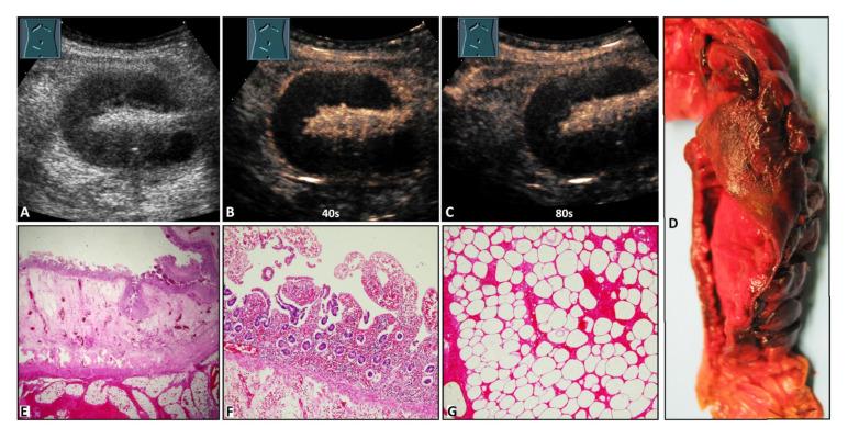
Figure 2.
A 42-year-old male patient with acute peritoneal pain in the right lower abdomen and a palpable subcutaneously located tumor in the form of a Spieghel’s hernia. (A) B-mode ultrasound shows a standing loop of small bowel. (B,C) Contrast-enhanced ultrasound shows absent enhancement of the bowel wall after 40 s and 80 s. (D) Macroscopic evaluation shows a color change to deep red in the mucosa in an 8 cm long segment. (E) Cross section of the small intestine with luminal mucosa, edematous submucosa, muscularis, and hemorrhagic submucosa (from top to bottom). The mucosa shows incomplete ischemic necrosis with vital crypt epithelium (1.25× magnification). (F) Small intestinal mucosa with incomplete ischemic necrosis with vital crypt epithelium (10× magnification). (G) Necrotic mesenteric fat tissue with missing nuclei and hemorrhages (10× magnification).

